Where were X-rays discovered, and by whom?
On 8 November 1895, German physicist Wilhelm Conrad Röntgen discovered X-rays at the University of Würzburg’s Physics Institute. He was investigating electrical conduction in gases. The radiation he observed could pass through materials that blocked visible light. He called it X-rays.
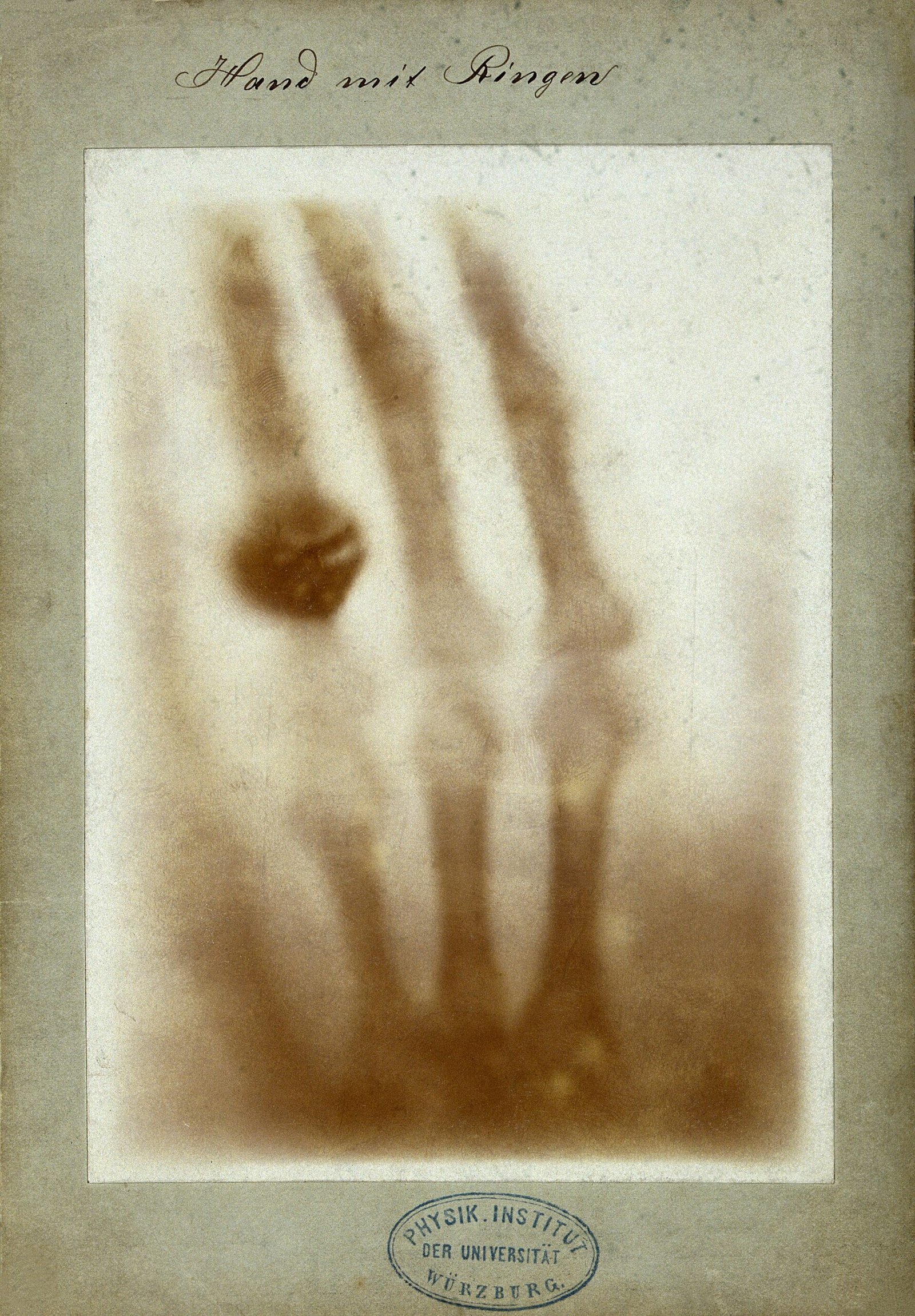
If you picture the discovery through its famous hand image, start with the laboratory instead. The discovery came in November. The radiograph of Anna Bertha Röntgen’s left hand followed on 22 December 1895, according to the Science Museum’s dating. Observation and the familiar image belong to separate moments.
Röntgen published his first paper announcing the new kind of rays on 28 December 1895. On 5 January 1896, the Viennese newspaper Die Presse reported the discovery. These dates mark the movement of X-rays from a laboratory observation into scientific publication and newspaper coverage.
What made X-ray vision possible?
X-rays are electromagnetic radiation, like visible light. Their higher energy allows them to pass through many objects, including the human body. Röntgen’s observation concerned radiation that penetrated substances opaque to visible light. That ability became the basis of medical X-ray imaging.
The resulting image depends on differences in absorption. Calcium in bones absorbs more X-rays than many surrounding soft tissues. Bones therefore look whiter on standard radiographs. If you have seen a dental X-ray, you have encountered an everyday application of this same radiation: radiography can identify some dental problems.
Medical radiography also helps detect bone fractures, pneumonia, and foreign objects inside the body. These uses connect Röntgen’s discovery to practical questions about injury and illness.
The hand image and the name Röntgen rays
The famous early radiograph shows Anna Bertha Röntgen’s left hand with a ring. The Science Museum dates its creation to 22 December 1895. The bones and ring make this particular hand image a concrete record of the new radiation’s imaging possibilities.
Another hand became part of the discovery’s public presentation. At the Würzburg Physical and Medical Society on 23 January 1896, anatomist Albert von Kölliker had his hand imaged. He proposed naming the radiation Röntgen rays. The proposal linked the rays directly to their discoverer.
Röntgen received the first Nobel Prize in Physics on 10 December 1901 for his discovery. The award followed the laboratory observation, the first paper, newspaper coverage, and the Würzburg presentation. Together, these events show how the discovery gained scientific recognition.
How does an X-ray machine produce radiation?
An X-ray tube is a vacuum tube. Electrons leave a heated cathode and accelerate under high voltage towards a metal anode target. Their collision with the target produces X-rays. This gives you a way to connect the machine’s electrical operation with the radiation used for imaging.
One production process is called bremsstrahlung, or braking radiation. Electrons striking a target such as tungsten slow down. As they lose energy, they release part of it as X-ray radiation. The term describes the slowing of the electrons at the target.
A second process produces characteristic X-rays. A vacancy in an atom’s inner electron shell is filled by another electron. The transition releases a photon with a particular energy. Both this process and braking radiation explain how interactions involving electrons can generate X-rays.
The tube voltage limits the energy of the photons produced. A tube operating at 80 kilovolts cannot generate photons with energies above 80 kiloelectronvolts. A later milestone in tube development was William David Coolidge’s paper, published in Physical Review on 1 December 1913.
From the laboratory to hospital medicine
By the 1930s, most hospitals in the United Kingdom had dedicated X-ray departments and equipment. The radiation discovered in Würzburg had become part of organised hospital care. Dedicated departments placed X-ray examination within the hospital’s own facilities.
Medical X-ray imaging now includes conventional radiography, computed tomography, fluoroscopy, and mammography. All use ionising X-ray radiation to form images. The names describe different imaging methods, but each belongs to the medical use of the radiation Röntgen discovered.
X-rays also have a treatment role. Radiotherapy uses them to target cancer cells. This application sits alongside diagnostic imaging: the same type of radiation can help produce an image or be used in cancer treatment.
X-ray technicians and wartime imaging
During the First World War, Marie Curie used donated automobiles to help establish mobile X-ray services. Her network included 20 mobile stations and 200 fixed stations.
Irène Joliot-Curie operated X-ray equipment on the Belgian front at the age of 18. She also taught nurses how to use it. Her work shows that wartime radiology involved both operating machines and passing practical skills to other medical workers.
The wartime stations and the dedicated hospital departments of the 1930s illustrate different settings for X-ray work. Curie’s network combined mobile and fixed equipment. British hospitals later housed their own departments. In both settings, medical imaging depended on equipment and people able to operate it.
What is X-ray crystallography?
X-rays also became a tool for studying crystals. Max von Laue’s work on X-ray diffraction in crystals was published in 1912. It established the electromagnetic nature of the rays. He received the 1914 Nobel Prize in Physics for the discovery of their diffraction by crystals.
William Henry Bragg and his son, William Lawrence Bragg, shared the 1915 Nobel Prize in Physics. Their recognised contribution concerned the analysis of crystal structures using X-rays. The radiation that could reveal structures inside a hand also supported investigations of crystal structure.
Why does radiation protection matter?
X-rays are ionising radiation and can damage DNA. That damage can contribute to genetic changes that lead to cancer. Their medical value therefore comes with a need to assess the exposure involved in an examination.
The FDA identifies two central protection principles. The expected benefit to the patient should outweigh the harm. The examination should also use the lowest dose that provides an image adequate for diagnosis. If you connect modern imaging with Röntgen’s discovery, these principles belong in that history alongside the images and machines.
Timeline
- 8 November 1895: Röntgen discovers X-rays in his Würzburg laboratory.
- 22 December 1895: Anna Bertha Röntgen’s famous hand radiograph is made, according to the Science Museum’s dating.
- 28 December 1895: Röntgen publishes his first paper announcing the discovery.
- 5 January 1896: Die Presse reports the discovery.
- 23 January 1896: Kölliker proposes the name Röntgen rays at the Würzburg presentation.
- 10 December 1901: Röntgen receives the first Nobel Prize in Physics.
- 1912: Laue’s work on X-ray diffraction in crystals is published.
- 1 December 1913: Coolidge’s paper describing his X-ray tube appears in Physical Review.
- 1914: Laue receives the Nobel Prize in Physics.
- First World War: Marie Curie establishes a network of mobile and fixed X-ray stations; Irène operates X-ray equipment and teaches nurses to use it.
- 1915: William Henry Bragg and William Lawrence Bragg share the Nobel Prize in Physics.
- 1930s: Most British hospitals have dedicated X-ray departments.
- 1962: A rocket experiment discovers Scorpius X-1.
Details you won’t find in guidebooks
The Science Museum’s familiar hand image is a photographic copy. Its collection record locates the original radiograph at the Deutsches Museum in Munich. When you look at this museum record, the distinction between the original image and its later copy matters.
Röntgen donated his Nobel Prize money of 50,000 kronor to the University of Würzburg. He did not apply for a patent for the discovery. He also did not deliver a Nobel lecture. These details accompany the more familiar record of his 1901 award.
From medical images to X-ray astronomy
Earth’s atmosphere absorbs most X-rays arriving from space. Astronomical detectors therefore need to be carried above the atmosphere. Observing these rays requires a different setting from a hospital examination, where a tube produces the radiation used to make an image.
In 1962, a rocket experiment by Riccardo Giacconi and his colleagues discovered Scorpius X-1. It is recognised as the first X-ray source discovered beyond the Solar System. X-rays had become a means of investigating both structures on Earth and sources in space.
FAQ
Where were X-rays discovered, and by whom?
German physicist Wilhelm Conrad Röntgen discovered X-rays on 8 November 1895 at the University of Würzburg’s Physics Institute while investigating electrical conduction in gases.
Whose hand appears in the famous early X-ray?
The image shows Anna Bertha Röntgen’s left hand with a ring. The Science Museum dates the radiograph to 22 December 1895.
Why do bones look white on an X-ray?
Calcium in bones absorbs more X-rays than many surrounding soft tissues, making bones appear whiter on standard radiographs.
How did X-rays lead to crystallography?
Max von Laue’s published work on crystal diffraction in 1912 established the electromagnetic nature of X-rays. William Henry Bragg and William Lawrence Bragg later shared the 1915 Nobel Prize in Physics for their contributions to analysing crystal structures with X-rays.
Sources
- University of Würzburg Archives — Wilhelm Conrad Röntgen uni-wuerzburg.de
- Röntgen Memorial Site Würzburg — Wilhelm Röntgen wilhelmconradroentgen.de
- Science Museum Group — Röntgen’s Hand Radiograph collection.sciencemuseumgroup.org.uk
- NIH–NIBIB — X-Rays nibib.nih.gov
- Wikipedia — X-ray en.wikipedia.org
- ARPANSA — X-rays arpansa.gov.au
- Physical Review — A Powerful Röntgen Ray Tube with a Pure Electron Discharge journals.aps.org
- FDA — Medical X-ray Imaging fda.gov
- American Institute of Physics — Marie Curie: Help for the Wounded history.aip.org
- NobelPrize.org — Irène Joliot-Curie nobelprize.org
- International Union of Crystallography — Nobel Prize in Physics 1914 iucr.org
- NobelPrize.org — William Bragg: Facts nobelprize.org
- National Cancer Institute — Radiation cancer.gov
Image: First medical X-ray by Wilhelm Röntgen of his wife Anna Bertha Ludwig’s hand – 18951222.jpg — Wilhelm Röntgen, Public domain